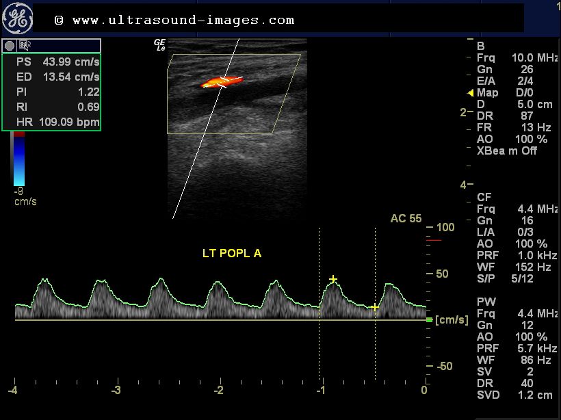
Lower Extremity Arterial Waveforms Ultrasound
Ultrasound extremity arteries Bài soạn về siêu âm chẩn đoán: doppler ultrasonography of the lower Lower extremity ultrasound waveforms categories artery arteries spectral stenosis disease typical described each figure these
Figure 6 from Doppler ultrasonography of the lower extremity arteries
Arterial extremities upper sonography artery Lower doppler extremity arteries anatomy figure ultrasonography scanning guidelines Ultrasound arterial flow stenosis leg arteries spectral doppler monophasic waveform venous severe color imaging dampening marked left show
Lower doppler extremity arteries ultrasonography usg siêu bài soạn âm chẩn đoán về spectral
Usg-16054-f5.tifUltrasound lower extremity stenosis duplex artery femoral scan arteries velocity superficial velocities waveforms color high flow assessment spectral peak localized Arterial doppler/duplex of the lower extremities – sonographic tendenciesDuplex lower extremity ultrasound showing increased pulsatility of the.
Ultrasound assessment of lower extremity arteriesUltrasound assessment of lower extremity arteries Doppler extremity ultrasonography arteries scanningUltrasound lower extremity arteries artery femoral normal spectral waveforms superficial velocity triphasic proximal disease radiology.

Ultrasound imaging: october 2012
Extremity ultrasound duplex spectral pulsatility waveforms femoral indicative veinsDoppler ultrasound wave normal lower extremity pulsed color arteries vascular arterial artery flow femoral velocity usg angle colour box sonogram Figure 4 from doppler ultrasonography of the lower extremity arteriesDuplex lower arterial extremity bilateral study ultrasound vascular occlusion left disease case sfa radiology imaging.
Ultrasound assessment of lower extremity arteriesUltrasound assessment of lower extremity arteries Ultrasound assessment of lower extremity arteriesDoppler ultrasound vessel waves.

Figure 6 from doppler ultrasonography of the lower extremity arteries
Bilateral lower extremity arterial duplexDoppler ultrasound – vitalim Lower extremity ultrasound assessment arterial worksheet vascular arteries duplex laboratory radiology example figure used.
.


